
Syllidae marine worms
from the Iberian Peninsula

Introduction
Syllidae family compiles the group of polychaetes characterized by a specialization in their foregut named as proventricle (1). This conspicuous structure, found right after the pharynx, is the autapomorphy that sets the basis for the complex systematics and taxonomy that these animals face, taxa composed approximately of 74 genera and more than 900 species (2, 3).

DISTRIBUTION AND HABITAT
Virtually, every coast of any nature is home to a multitude of syllids. They are usually present among algae, rhizomes of marine phanerogams, sands and sediments of all kinds. However, even though they are extraordinarily abundant in littoral samples, they are somewhat scarcer at great depths (4). They were found to inhabit many areas in the Mediterranean Sea, mainly in the Iberian peninsula (Fig. 1), where we can find one of the greatest diversity on syllids (5).
.png)
Figure 1. Syllidae distribution through the Iberian Peninsula, based on data collected from GBIF. Original work
MORPHOLOGY
Syllids are medium-sized annelids, with a subcylindrical body divided into homonymous segments. Their anatomy can be divided into 3 main sections: the anterior part, containing the prostomium, with the antennae, palps and eyes, the pharynx, with its pharyngeal tooth, the peristomium and the beginning of the proventricle. The middle part consists mainly of the rest of the proventricle and body, and the posterior part includes the progressive slimmer end of the body. These sections are also well differentiated as they present differences in their cirrus, parapodial lobes, chaetae and aciculae (Fig. 2).

Figure 2. Diagram of the main morphological characters of the Syllidae family, classified according to the area of the body in which they are located. Illustration of Syllis sp. [Note: the delimitation of the middle and posterior part is merely illustrative. To determine in detail the limit or transition of both parts in each species, it is necessary to morphologically differentiate the middle setae from the posterior chaetae]. Original work
In order to describe a species of Syllidae, chaetae are essential (Figs. 3, 4). They can differ throughout the body, in shape and size, being compound (divided into shaft and blade) (Fig. 3A, 3B) or simple (lacking this trait) (Fig. 3C, 3D, 3E, 3F). Aciculae provide support to parapodia. Its morphology also helps with the description of species (Fig. 4).
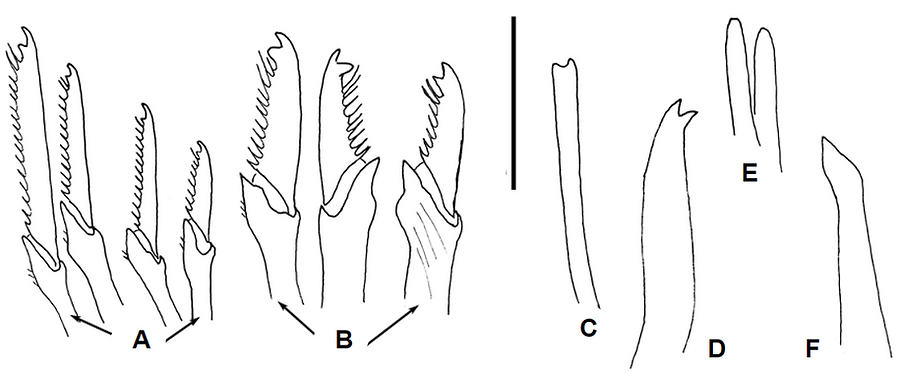
Figure 3. Compound chaetae detailed in B and C, simple chaetae detailed in D and E, aciculae detailed in F and G. Scale: A, 0.1 mm; B-G, 20 µm. (7)

Figure 4. Detailed line drawings of different types of chaetae: C. Spiniger chaetae, D., E. Falciger chaetae, F. Claw-shaped chaetae, G. Ypsiloid chaetae, H., I., J., K. Simple chaetae. Abbreviations: bd, blade; sf, shaft (3).
SYSTEMATICS
Syllidae family is divided into 5 subfamilies that mainly differ in several characters of appendages, palps and chaetae, all easily to distinguish morphologically (Fig. 5).

Figure 5. Phylogenetic tree of the 5 subfamilies of Syllidae. Drawings from top to bottom: Streptospinigera niuqtuut (7), Sphaerosyllis parabulbosa (9), Autolytus labordai (8), Odontosyllis aracaensis (9) and Syllis jorgei (10).

Subfamilies


Bibliography
Syllidae colour image © Alexander Semenov, Moscow State University’s White Sea Biological Station, 2020.
1. Franke, HD. (1983). Wilhelm Roux's Archives of Developmental Biology, 192. 2. San Martín, G. and Worsfold, TM. (2015). Zookeys, 488. 3. Álvarez-Campos, P. (2016), Doctoral dissertation, Universidad Autónoma de Madrid. 4. San Martín, G. (2003). Museo Nacional de Ciencias Naturales, CSIC, 21. 5. Musco, L. and Giangrande, A. (2005). Marine Ecology Progress Series, 304. 6. Álvarez-Campos et al. (2015). Zootaxa, 4052. 7. Olivier et al. (2013). Polar biology, 36. 8. San Martín and López (2002). Sarsia, 87(2). 9. Fukuda et al., 2013. Zootaxa, 3609(2). 10. San Martín and López, 2000. Cahiers de Biologie Marine, 41(4).

More information
Click on a bubble to know more!
.png)
GLOBAL DATA
.png)
SEQUENCES
.png)
TAXONOMY
.png)
TRAITS
.png)
DISTRIBUTION